Parasite Portfolio

Welcome to the Parasite Portfolio, a comprehensive study guide for our parasitology part of the Lab class. This portfolio brings together essential terms, diagnostic procedures, lifecycle details, and visuals of various parasites we saw in lab.
I was a software developer for 6 years and I thought it would be fun to create a little website for this assignment.

Common Terms

Lab Equipment & Procedures

A compound light microscope consists of 2 separate lens systems: the ocular (9) and the objective (3-5).

  • Ocular lens - magnification x10
  • Objective lens - magnification x4 (scanning), x10 (low power), x40 (high dry), x100 (oil immersion)

Total magnification = 10 (ocular) x N (objective) = 10N

Microscope parts

A device that applies a centrifugal (rotation) force to separate various components of a liquid based on their density.

Example:
Blood = RBC + WBC + plasma/serum Urine = sediment + supernatant Fecal flotation = meniscus + solution + sediment

Centrifuge parts
Centrifuge parts

Source

Battlefield Method to scan a slide

Source - Lab Lecture Presentation

The number of ova or parasites is the number viewed on the entire slide for each parasite reported

Ova & Oocyte Reporting

Source - Lab Lecture Presentation

Materials:

  1. Glass slide
  2. Glass coverslips
  3. Wooden applicator stick
  4. Water or saline
  5. Waxed paper cups

Procedure:

  1. Dip the applicator stick into the feces (only a small amount should adhere to the stick).
  2. Place a drop of saline on a slide.
  3. Mix the feces with the saline to produce a homogeneous emulsion that is clear enough to read newsprint through (a common mistake is to make the smear too thick).
  4. Place the coverslip over the emulsion.
  5. Examine the slide.

Materials:

  1. Glass slide
  2. Glass coverslips
  3. Wooden applicator stick
  4. Paper cup
  5. Waxed paper
  6. Gauze squares of metal screen tea strainer
  7. Funnel
  8. Conical tube
  9. Sugar flotation solution
  10. Scale

Procedure:

  1. Place approx. 5 g of feces in the paper cup.
  2. Add 30 ml of flotation solution.
  3. With the applicator stick, mix the feces to an evenly suspended emulsion.
  4. Using the strainer, pour the liquid into the tube enough to create a meniscus (dome).
  5. Place a coverslip on top of the tube.
  6. Leave the coverslip for 15 minutes.
  7. Pick the coverslip and place it on the glass slide with the fluid down.
  8. Examine the slide.
Fecal Flotation Containers

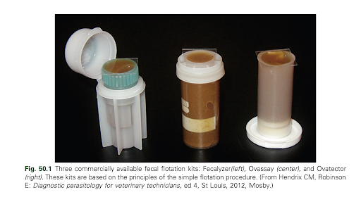
Source - Hickie, J., & Blanchard, L. (2024). Fig. 50.1. Laboratory Procedures for Veterinary Technicians (7th ed., p. 323). Elsevier Canada.

Materials:

  1. Glass slide
  2. Glass coverslips
  3. Wooden applicator stick
  4. Paper cup
  5. Gauze squares of metal screen tea strainer
  6. Funnel
  7. Conical tube
  8. Sugar flotation solution
  9. Scale

Procedure:

  1. Prepare a fecal emulsion using 5 g of feces and 30 mL of the flotation solution.
  2. Using the strainer, pour the liquid into the centrifuge tube until it’s about ⅔ full.
  3. Place a waxed paper on top of the tube.
  4. Place the tubes in the centrifuge buckets, and balance them with the tube with equal weight of the sample or water.
  5. Centrifuge the tubes for 5 minutes at approximately 1300 rpm.
  6. Remove the coverslips from the tubes by lifting straight up, and then place it on a slide.
  7. Examine the slide.

Materials:

  1. Glass slide
  2. Glass coverslips
  3. Wooden applicator stick
  4. Paper cup
  5. Gauze squares of metal screen tea strainer
  6. Funnel
  7. Conical tube
  8. Sugar flotation solution
  9. Scale
  10. Disposable pipettes

Procedure:

  1. Prepare a fecal emulsion using 5 g of feces and 30 mL of the flotation solution.
  2. Using the strainer, pour the liquid into the centrifuge tube until it’s about ⅔ full.
  3. Place a waxed paper on top of the tube.
  4. Place the tubes in the centrifuge buckets, and balance them with the tube with equal weight of the sample or water.
  5. Centrifuge the tubes for 5 minutes approximately 1300 rpm.
  6. Use a pipette to carefully remove the solution, leaving the supernatant at the bottom.
  7. Use a pipette to transfer the sediment to a slide.
  8. Place a coverslip over the sediment.
  9. Examine the slide.

Materials:

  1. Glass slide
  2. Glass coverslips
  3. Wooden applicator stick
  4. Baermann apparatus (tube stand, funnel, rubber tubing, clamp, and wire screen)
  5. Water
  6. Funnel
  7. Gauze squares of metal screen tea strainer
  8. Conical tube
  9. Scale
  10. Disposable pipettes

Procedure:

  1. Construct the Baermann apparatus by putting the funel to a tube stand.
  2. Attach 3 to 4 inches of rubber tubing to the narrow portion of the funnel and ensure a good seal.
  3. Put the tea strainer on top of the funel.
  4. Place pinch clamps at the end of the rubber tubing and check for a tight seal using water.
  5. Wrap 30 g. of feces in several layers of cloth like a teabag and place the fecal pouch in the funnel.
  6. Fill the funnel with warm water (not hot) above the fecal sample.
  7. Allow the apparatus to remain undisturbed for 24 hours.
  8. After the time has passed, collect approx. 2 ml of the fluid from the rubber tubing and transfer it to a centrifuge tube.
  9. Use a pipette to transfer the supernatant to a slide.
  10. Place the coverslip over the emulsion.
  11. Examine the slide.

Materials:

  1. McMaster slides
  2. Paper cup
  3. Scale
  4. Sugar flotation solution
  5. Wooden applicator stick
  6. Disposable pipettes
  7. Gauze squares of metal screen tea strainer

Procedure:

  1. Prepare a fecal emulsion using 4 g of feces with 56 mL of the flotation solution.
  2. Strain the emulsion with a tea strainer to remove any large pieces of debri.
  3. use a filter pipette to transfer material to the counting chamber.
  4. Allow the slide to sit undisturbed for 10 minutes.
  5. Using the 10× objective lens, focus on the etched grid of the McMaster slide. Count all eggs or oocysts in the six columns of the etched square. Keep a separate count for each species of parasite observed.
  6. Multiply the number of eggs or oocysts counted by the appropriate dilution factor based on the number of squares counted. Record the results as eggs per gram (epg) of feces. The volume under the etched area is 0.15 mL.
  7. Use the formula: (chamber 1 + chamber 2) * 50 = eggs per gram (EPG)

Parasite Classification

Parasites Classification

Endoparasites

Click to view details

Endoparasites

Click to view details

Ectoparasites

Click to view details

Ectoparasites

Click to view details